
First, the paper
EKG paper runs at a standard speed, and the grid is a clock:
|
On the paper |
Equals |
|
1 small box |
0.04 seconds |
|
1 large box (5 small) |
0.20 seconds |
|
5 large boxes |
1 second |
|
30 large boxes |
6 seconds |
Most rhythm strips are printed as 6 seconds, which matters for the fastest way to count a rate.
The five steps
Same order, every strip, whether it looks obvious or not.
1. Rate
Normal is 60 to 100 beats per minute.
Two ways to count it:
6-second method: count QRS complexes in 6 seconds × 10
300 method: 300 ÷ number of large boxes between two R waves
The 6-second method works on any rhythm, regular or not, and it is the one to use when the rhythm is irregular. The 300 method is faster but only reliable when the rhythm is regular.
Worked example: 8 QRS complexes on a 6-second strip means 8 × 10 = 80 bpm.
2. Rhythm
Regular or irregular? March out the R-to-R intervals — with calipers, or by marking two R waves on a piece of paper and sliding it along the strip.
If the R-R intervals are equal, the rhythm is regular. If they vary with no pattern at all, that is described as irregularly irregular, which is a phrase worth knowing because it points strongly toward one rhythm in particular.
3. P waves
Four questions about them:
- Is there a P wave before every QRS?
- Is there a QRS after every P wave?
- Are the P waves upright and uniform in shape?
- Is there exactly one P wave per QRS?
Absent P waves, multiple P waves per QRS, or P waves marching independently of the QRS all point somewhere specific.
4. PR interval
Measure from the start of the P wave to the start of the QRS.
Normal: 0.12 – 0.20 seconds (3 to 5 small boxes)
Longer than 0.20 and constant, or longer and getting progressively longer, or varying with no pattern — each of those means something different, and this single measurement distinguishes the heart blocks from one another.
5. QRS duration
Measure from the start to the end of the QRS complex.
Normal: less than 0.12 seconds (under 3 small boxes)
A narrow QRS means the impulse came from above the ventricles. A wide QRS means it originated in the ventricles or took an abnormal path through them. That one distinction separates a large group of rhythms from another.
What the answers tell you
Once you have five measurements, the rhythm follows from them:
|
Rhythm |
Rate |
Regular? |
P waves |
QRS |
|
Normal sinus |
60–100 |
Regular |
One before each QRS |
Narrow |
|
Sinus bradycardia |
Under 60 |
Regular |
One before each QRS |
Narrow |
|
Sinus tachycardia |
Over 100 |
Regular |
One before each QRS |
Narrow |
|
Atrial fibrillation |
Varies |
Irregularly irregular |
Absent — wavy baseline |
Narrow |
|
Atrial flutter |
Varies |
Often regular |
Sawtooth flutter waves |
Narrow |
|
SVT |
150–250 |
Regular |
Often hidden in the T wave |
Narrow |
|
Ventricular tachycardia |
Over 100 |
Regular |
Absent |
Wide |
|
Ventricular fibrillation |
None |
Chaotic |
Absent |
No true QRS |
|
Asystole |
None |
— |
Absent |
Absent |
The heart blocks
All four are distinguished almost entirely by what the PR interval is doing:
|
Block |
What the PR interval does |
|
First degree |
Constant, but longer than 0.20 seconds. Every P wave gets a QRS. |
|
Second degree, Type I (Wenckebach) |
Gets progressively longer, beat after beat, until one QRS is dropped entirely — then the cycle restarts. |
|
Second degree, Type II |
Stays constant, but some P waves are not followed by a QRS at all. |
|
Third degree (complete) |
No relationship at all. P waves and QRS complexes march independently of each other. |
Type I is generally the more benign of the two second-degree blocks. Type II is the one that worries people, because it can progress to complete heart block.
The rhythms that mean act now
Some rhythms are interpretation questions. Some are emergencies.
- Ventricular fibrillation — no effective output, no pulse
- Pulseless ventricular tachycardia — an organized-looking rhythm with no pulse
- Asystole — no electrical activity
- Pulseless electrical activity — a rhythm on the monitor with no pulse at the patient
That last one is the reason for the single most important rule in this whole article:
Treat the patient, not the monitor. Always check a pulse. A monitor showing a perfect rhythm on a patient with no output is still an emergency.
And the opposite happens too. A flat line on the monitor with an awake, talking patient is almost always a lead that has come off. Look at the patient before you look at the strip.
How to actually get good at this
Repetition, in mixed order. Nothing else works as well.
- Use the same five steps on every strip, including the ones you think you recognize instantly
- Say your findings out loud — "rate 80, regular, P before each QRS, PR 0.16, QRS narrow" — before naming anything
- Practice strips grouped by rhythm type teach you very little; mixed strips teach you everything
- Work through dozens rather than a handful. Recognition is a volume effect.
- When you get one wrong, work out which of the five steps you got wrong, not just which rhythm it was
Free EKG cheat sheet
We made a one-page printable reference: the five steps, the normal intervals, the paper measurements, and a rhythm criteria table. Designed to fit in a badge holder or a clipboard.
Free, no purchase required.
Where to go from here
The method is the easy part. Getting it to the point where it is automatic under pressure takes practice strips, and a lot of them.
Our EKG & Dysrhythmia Interpretation Guide is 111 pages covering the scan method in depth, every rhythm and block with what causes it and what it means for the patient, and 65 practice strips — drills after each module plus a 35-strip final exam. The adult ACLS algorithms are included for reference. Written by a critical care nurse with over 15 years at the bedside.
Written by Jennifer Cheung, MSN, RN, CCRN. This article is educational and does not replace clinical training, your institution's protocols, or current resuscitation guidelines. Always follow your facility's procedures and escalate according to policy.
Featured Products
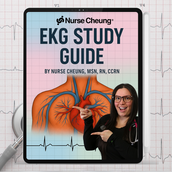
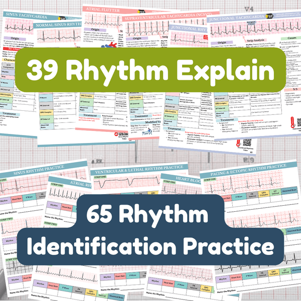
EKG & Dysrhythmia Interpretation Guide
111 Pages · 65 Practice Strips · ECG Rhythms & ACLS
Stop memorizing rhythm names and start reading the electrical story behind them. This 111-page EKG guide (also written ECG) teaches a repeatable method for reading any strip: rate, rhythm, P...

Nursing School Survival Bundle
Phase I & Phase II · 185 Pages
Both bootcamps together cover the whole path from application to the end of your first semester. Phase I gets you in and ready. Phase II gets you through.